Synthesis and characterization of block copolymers and NPs
The synthetic routes were minutely depicted in Fig. S1. Initially, employing a classical one-pot method [36], the diblock three-component copolymer with tunable stability, poly(cyclobutane−1,2,3,4-tetracarboxylic dianhydride-co-hydroxyethyl disulfide)-polyethylene glycol (mPEG5K–p(CBDA-co-BiBi)-mPEG5K, abbreviated as ssP-COOH), was synthesized, using 2-hydroxyethyl disulfide (BiBi) monomer and cyclobutane−1,2,3,4-tetracarboxylic dianhydride (CBDA) molecular synthesis as functional skeletons, as well as the introduction of mPEG5k-NH2 chain as hydrophilic fragment (Fig. S1). Based on the nucleophilic reactions, thus, the as-prepared ssP-COOH copolymer displayed numerous disulfide bonds (-S-S-) and pendent pairwise carboxylic acid (-COOH) groups, providing a foundation for creating diverse copolymers. Subsequently, an standard EDC/HOBT-catalyzed amidation reaction was performed to couple the side carboxylic acid group with ethylenediamine-monoboc (EDA-Boc), and then the modified copolymer was named as ssP-EDA-Boc copolymer. The next work was a simple in-situ Boc-deprotection reaction using trifluoroacetic acid (TFA) solution to form an amino-modified polymer (ssP-EDA copolymer). The chemical structures of these block copolymers were characterized through 1H nuclear magnetic resonance (1H NMR) spectroscopy (Fig. S2) and fourier transform infrared (FTIR) spectroscopy (Fig. S3) and UV-vis spectra (Fig. S4), confirming their successful synthesis.
Furthermore, it is well known that, in aqueous solution, amphiphilic block copolymers can spontaneously drive the formation of diverse nanoparticles (such as spheres, cylinders and lamellaes) through self-assembly approach [36, 37]. Therefore, the distinct NPs, including ssP-COOH, ssP-EDA-Boc, and ssP-EDA, were fabricated in mild solution. The diameter of these NPs decreased progressively from 197.6 nm to 146.1 nm and then to 108.1 nm, as analyzed by DLS, due to the gradual modification of copolymer (Fig. S5a, S5c), mainly probably due to the increased hydrophobic capacity and electrostatic adsorption effect. Moreover, variations in zeta potential further proved the success of grooming (Fig. S5b).
To enhance the strong active metal (e.g., Fe3+) carrying capacity of block copolymers, we incorporated a polyphenol-like structure with acid-responsive Schiff-base bonding, utilizing the 3,4,5-trihydroxybenzaldehyde (tHB) monomer. A new peak appeared around 7.9–6.7 ppm in the 1H NMR spectrum was attributed to the benzene ring structure, indicating the successful preparation of tHB-decorated block copolymer (ssP-tHB) (Fig. S2). Besides, the presence of v (C = N) azomethine stretching vibrations was confirmed by the detection of absorption peaks at 1720 cm − 1 for tHB, which indicates the establishment of Schiff base linkage [19].
Next, the FDA-approved doxorubicin (DOX), as a hydrophobic broad spectrum anti-cancer drug, often lacks bio-targeting, leading to cellular resistance and limiting its effectiveness in cancer treatment [19, 38]. To that end, using nano-precipitation, the ssP-tHB block copolymer created square nanoparticles that encapsulated DOX through hydrophobic interactions. These were then enhanced with polyphenol-iron coordination to form ssP-tHB@Fe/DOX (Fig. 1a), which boasts strong stability, minimal drug leakage, and high iron ion-carrying capacity.
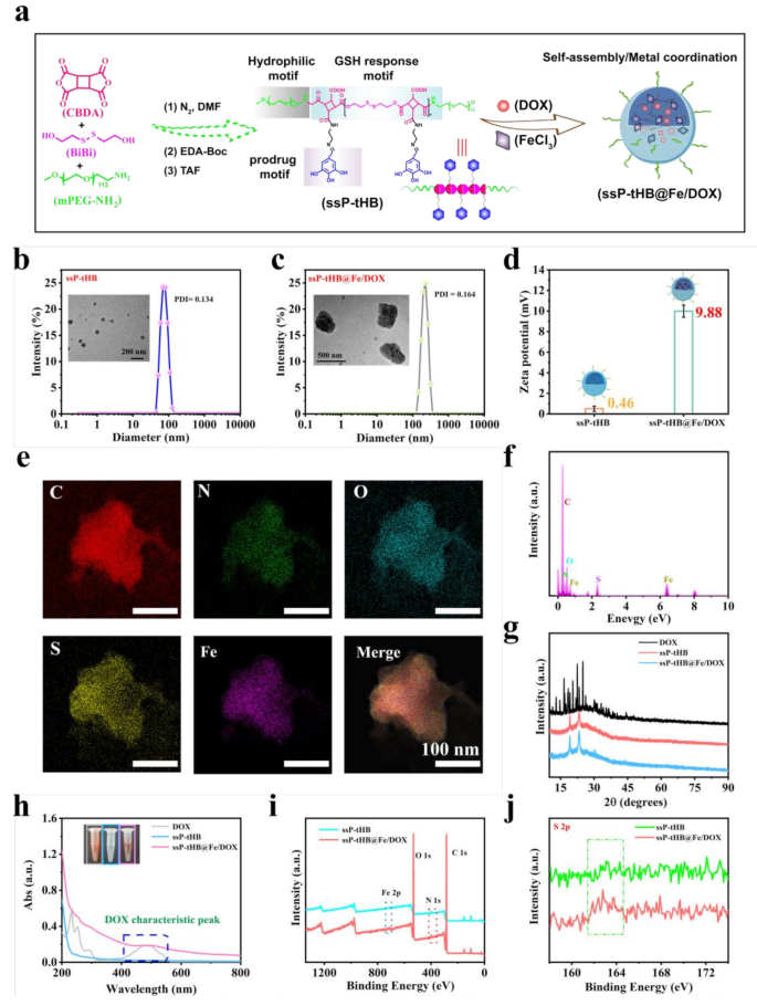
Preparation and characterization of ssP-tHB@Fe/DOX. (a) Synthesis routes of different nanoparticles. (b, c) Hydrodynamic sizes and TEM images of ssP-tHB (b), and ssP-tHB@Fe/DOX (c). (d) Zeta potential changes of nanoparticles (n = 5). (e) EDS elemental mapping images of ssP-tHB@Fe/DOX. (f) EDS elements of ssP-tHB@Fe/DOX. (g) Powder XRD patterns of free DOX, ssP-tHB, and ssP-tHB@Fe/DOX. (h) UV-vis absorption spectra of free DOX, ssP-tHB, and ssP-tHB@Fe/DOX. Inset: The corresponding photos of samples. (i) XPS spectra of ssP-tHB and ssP-tHB@Fe/DOX. (j) High-resolution S 2p XPS spectra of ssP-tHB and ssP-tHB@Fe/DOX
After that, the composition and structure of ssP-tHB and Fe/DOX-coloaded ssP-tHB were minutely characterized. By DLS testing, in aqueous solution, the self-assembled ssP-tHB block copolymer prominently formed spherical nanoparticles with a hydrodynamic particle size of 147 nm and a polydispersity index (PDI) of 0.134 (Fig. 1b). Fe/DOX-loaded NPs have a larger hydrodynamic diameter of approximately 220 nm and a PDI of 0.164 compared to blank NPs, demonstrating successful payload incorporation (Fig. 1c).
In TEM images, ssP-tHB@Fe/DOX in rough tablet-like morphology with about 350 nm in length and 200 nm in width was showed, whereas blank NPs were uniformly spherical, measuring 50–60 nm (Fig. 1b), indicting that DOX loading and iron ions coordination can influence the morphology of the nanoassemblies. Furthermore, the self-assembled ssP-tHB solution shrunk in its dry state due to the dehydration effect [19, 20]. However, this similar phenomenon did not occur with ssP-tHB@Fe/DOX, primarily because iron-phenol crosslinking stabilized the nanoparticle structure.
The zeta potential of ssP-tHB decreased from 13.42 mV (ssP-EDA) to 0.46 mV, attributed to the negative charges of tHB pendant groups [39]. Meanwhile, a positively charged ssP-tHB@Fe/DOX (9.88 mV) was observed compared to blank NPs (ssP-tHB) (Fig. 1d), mainly due to the successful incorporation of the positively charged DOX drug and iron ions. As showed in UV-vis absorption spectra (Fig. 1h), ssP-tHB illustrated no obvious characteristic absorption peak. Conversely, the absorption peak at 410–550 nm, characteristic of ssP-tHB@Fe/DOX or DOX, indicates the successful incorporation of DOX into the nanoplatform.
To investigate the loading of DOX and iron ions, we subsequently conducted a series of experiments. In the study, through solution self-assembly, the anticancer drug DOX was encapsulated, followed by the introduction of trivalent iron ions for metal coordination to further prevent leakage of DOX and enhance the stability of NPs. Therefore, the prepared nanoparticles (ssP-tHB@DOX) had an average particle size of approximately 165 nm, with a PDI of 0.121 and a zeta potential of 4.56 mV, via DLS measurement (Fig. S6a), indicating that the hydrophobic chemotherapeutic drug DOX was primarily encapsulated within the hydrophobic region of block copolymers through self-assembly method. Furthermore, the UV-vis spectra of different samples were analyzed. As shown in Fig. S6b, after mixing free DOX with Fe3 + solution, the characteristic peak of DOX at about 480 nm did not fluctuate, indicating that the chelation between iron ions and DOX did not interfere with the characteristic absorption peak of DOX. Subsequently, when ssP-tHB@Fe/DOX was added in DMSO solution, it was clearly observed through digital photography that the red color of the DMSO solution became deeper compared to in aqueous solution, and the characteristic peaks of DOX also enhanced by UV-vis spectra (Fig. S6c), indicating that the DMSO solution effectively disrupted the polymer-based nanoparticles, promoting the release of DOX from the hydrophobic cavities of the copolymer. Furthermore, the DMSO-treated ssP-tHB@Fe/DOX was divided into two segments, with sizes ranging from 40 to 68 nm and 531–955 nm (Fig. S6d), further demonstrating that DMSO solution could effectively disrupt NPs stability. As a result, the weakening of DOX’s characteristic peaks after drug loading could be attributed to: (a) effective incorporation of the hydrophobic chemotherapeutic anticancer drug DOX into the polymer’s hydrophobic layer during the solution self-assembly process; (b) interference with DOX’s characteristic absorption peak due to π-π stacking interactions involving its large benzene rings [20, 39]. Simultaneously, Fe3 + mainly utilized the coordination between phenol group and iron ions to effectively carry iron ions on the copolymer surface.
According to the DOX concentration-dependent absorption standard curve, the content of DOX in ssP-tHB@Fe/DOX estimated to be 21.2% (Fig. S7). Moreover, the mass percentage of Fe in ssP-tHB@Fe/DOX about was 17.3% through ICP technology. The Fig. 1e showed the distribution of elements for ssP-tHB@Fe/DOX, and related elements such as C, N, O, S and Fe were evenly dispersed on its the surface, indicating the successful preparation of nanoparticles. In addition, the energy-dispersive X-ray (EDC) spectrum further corroborated this finding (Fig. 1f). A consequent X-ray photoelectron spectroscopy (XPS) characterization further confirmed the construction of the ssP-tHB@Fe/DOX (Fig. 1i). Meanwhile, the typical peak about at 163.8 eV indexed as S-S or S-C bonds [40], was observed in S sophisticated XPS spectra of ssP-tHB and ssP-tHB@Fe/DOX, effectively confirming the presence of nanoparticles responsive to GSH (Fig. 1j). Following this, we performed an XRD analysis on the crystal structures of ssP-tHB, ssP-tHB@Fe/DOX, and free DOX powder. The results indicated that both the solution self-assembled nanoparticles and the DOX drug exhibited an amorphous structure [39]. During self-assembly, the characteristic diffraction peaks at 10º−40º of DOX vanished compared to ssP-tHB or ssP-tHB@Fe/DOX (Fig. 1g), likely due to π-π stacking and hydrophobic interactions between them [39].
GSH-responsive degradation and catalytic performance
The favorable colloidal stability and appropriate size of nanoparticles are crucial factors for the accumulation in tumor regions enhanced by the permeability and retention (EPR) effect [19, 41]. We further investigated the stability of ssP-tHB@Fe/DOX in different solution (PBS, 10% FBS medium, and dH2O) (Fig. 2a). There were no significant changes in particle distribution across different media. Additionally, the hydrodynamic diameters of ssP-tHB@Fe/DOX remained constant over the seven-day tracking experiment (Fig. 2b). This establishes a foundation for using ssP-tHB@Fe/DOX in tumor therapy through caudal intravenous injection.
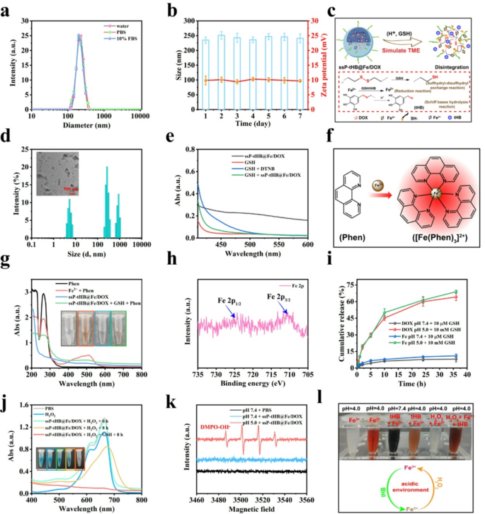
GSH responsive performance and ·OH generation assessments. (a) Hydrodynamic size of ssP-tHB@Fe/DOX in different media (water, PBS, and 10% FBS) (n = 5). (b) Changes of size and zeta potential of ssP-tHB@Fe/DOX in PBS for 7 days (n = 5). (c) The diagram of H+/GSH-emitted degradation and release of ssP-tHB@Fe/DOX. (d) Changes in hydrodynamic size of ssP-tHB@Fe/DOX in pH 5.0 + 10 mM GSH at 12 h. Inset: The corresponding TEM photo of samples. (e) UV-vis spectra for GSH level detection using DTNB as a probe in the presence of ssP-tHB@Fe/DOX. (f) Schematic illustration for Fe2+ detection using 1,10-Phenanthroline (Phen) probe. (g) UV-vis spectra for Fe2+ detection using 1,10-phenanthroline as a probe in different mixtures. Inset: The corresponding photographs of samples (from left to right). (h) High-resolution XPS spectrum of Fe 2p in ssP-tHB@Fe/DOX. (i) The release profile of DOX and Fe ions from ssP-tHB@Fe/DOX under different stimulation conditions (n = 5). (j) EPR spectra of DMPO-OH in aqueous solutions in different conditions. (k) UV-vis absorption spectra of regarding ssP-tHB@Fe/DOX-mediated MB degradation under different conditions. (l) Digital photos showing interactions between iron ions and tHB molecules under different conditions
Current research indicates that tumor regions display significant differences from normal tissues, such as elevated levels of GSH, an acidic environment, and various enzymes that promote tumor growth [16, 19]. Therefore, these tumor-specific nurturing microenvironments will enhance the effectiveness of intelligent nanotechnology delivery systems, particularly tumor-responsive drug release. Based on this, the nanoparticles containing S-S bonds and Fe3 + ions were assessed under GSH conditions (Fig. 2c). As depicted in Fig. 2d, ssP-tHB@Fe/DOX undergone severe disintegration at pH 5.0 with 10 mM GSH over 12 h, as observed through TEM imaging. DLS analysis revealed three distinct particle size signals for acid/GSH-treated ssP-tHB@Fe/DOX: small fragments (5–10 nm), larger particles (300–400 nm), and micron-sized particles. This suggests that acid and GSH conditions can cause the degradation of ssP-tHB@Fe/DOX. We then explored the capacity of GSH consumption of ssP-tHB@Fe/DOX or ssP-tHB through the 5,5’-dithiobis-(2-nitrobenzoic acid) (DTNB) chromogenic method (Fig. 2e, S8). Nanoparticles containing plentiful S-S bonds showed a robust GSH-depleting capability, relying mainly on the presence of nanoparticle-rich disulfide bonds and high-valence iron ions. So, these findings indicate that ssP-tHB@Fe/DOX has GSH-triggered ferroptosis-inducing potential for tumor treatment.
Consequently, the crucial role of iron in the induction of ferroptosis was examined. Therefore, the 1,10-phenanthrolline (Phen) probe was selected for detecting bivalent iron ions in ssP-tHB@Fe/DOX using UV-vis spectroscopy (Fig. 2f). The red flocculation was clearly showed when GSH treated ssP-tHB@Fe/DOX, indicating the production of Fe2 + by GSH reduction (Fig. 2g). The high-resolution XPS spectrum of Fe 2p revealed that Fe in ssP-tHB@Fe/DOX exists as both Fe(II) and Fe(III). Specifically, Fe 2p3/2 and Fe 2p1/2 peaks were observed at 711.4 eV and 727.5 eV [42], respectively (Fig. 2h), indicating tHB-induced generation of Fe2 + from ssP-tHB@Fe/DOX. Thus, we also investigated the chemical transformations of small molecule tHB and iron ions in various oxidation states across different media (Fig. 2l). The conversion of Fe3+/Fe2 + was tested by a 1,10-phenanthroline probe (Fig. 2f). Under acidic conditions (pH 5.5), tHB and trivalent iron ions exhibited a pale brown color, unlike the Fe(II) solution. At a pH of 5.5, the solution of Fe(II) exhibited a red color change upon the introduction of tHB, indicating the reduction of Fe(II) by tHB. Interestingly, the presence of H2O2 resulted in both Fe (II) and tHB small molecules displaying similar red colors. Thus, we can anticipate that the tHB-mediated Fe3+-to-Fe2 + conversion following ssP-tHB@Fe/DOX internalization by tumor cells will ensure continuous Fe2 + production, facilitating highly efficient Fenton reactions [43].
Next, given the high GSH expression in tumors that mediates the response to ssP-tHB@Fe/DOX, the drug release curves of the loaded DOX and iron ions were investigated (Fig. 2c). As shown in Fig. 2i, in pH 5.0 and 10 mM GSH (simulating tumor microenvironments), DOX from ssP-tHB@Fe/DOX displayed a continuous release action, achieving a cumulative release rate of 64.2% in 36 h, whereas only 11.3% of DOX was released during incubation in pH 7.4 and 10 µM GSH (simulating normal conditions) over the same time period. In addition, for iron ions in ssP-tHB@Fe/DOX, the analogous release profiles were collected by ICP testing, and approximately 68.9% of iron ions were released under simulated tumor conditions, while only 11.1% was released in the simulated normal conditions (Fig. 2i). Some factors contribute to these behaviors: (a) ssP-tHB@Fe/DOX contains -S-S- bonds that create the hydrophobic region leading to disassembly and the release of DOX and iron ions; (b) Fe (III) is reduced to Fe (II), weakening its coordination with polyphenol groups and accelerating release; (c) the Schiff base bonds attached to the tHB monomer detach in the presence of weak acid (pH 5.0), accelerating the disintegration of ssP-tHB@Fe/DOX by destroying the hydrophobic layer (Fig. 2c).
The release of iron ions from ssP-tHB@Fe/DOX induced the generation of highly toxic hydroxyl radicals (·OH) via the Fe-mediated Fenton reaction. Methylene blue (MB), used as a colorimetric substrate, was chosen for detection. As depicted in Fig. 2j, no obvious·OH formation was observed in a pH 7.4 environment with 10 mM H2O2 after 8 h, indicating the non-toxic nature of ssP-tHB@Fe/DOX prior to reaching the tumor site. However, under similar tumor conditions (pH 5.0 + 10 mM H2O2), ssP-tHB@Fe/DOX produced a considerable abundance of ·OH, resulting in the oxidation of MB. Amusingly, rich ·OH was detected when 10 mM GSH was present (resembling the tumor intracellular environment), indicating that GSH-treated Fe (III) produced active Fe (II) to generate ·OH, further demonstrating that ssP-tHB@Fe/DOX has tumor-activated therapeutic property. Electron spin resonance (ESR) spectroscopy, utilizing 5.5-dimethylpyrroline N-oxide (DMPO), was employed to determine ·OH levels (Fig. 2k). We found that pH 7.4 environment was not responsible for the production of ·OH, whereas iron-containing nanoparticles in pH 5.0 condition could generate abundant ·OH through the iron-based Fenton reaction in a manually simulated tumor microenvironments [44], indicating that its potential to induce ferroptosis in tumors.
Apoptosis and ferroptosis mechanism analysis in vitro
At present, treatment model of chemotherapy alone is prone to allow poor tumor efficacy and chemoresistance of tumor cells, thereby resulting in the failure of tumor treatment or tumor progression. Therefore, non-apoptotic death property, namely ferroptosis, was successfully combined to coordinate tumor therapy, in this study (Fig. 3h). It is well known that iron accumulation and subsequent lipid peroxidation (LPO) play crucial roles in triggering and accelerating ferroptosis. Therefore, the molecular pathways and signaling mechanisms involved in iron homeostasis and LPO play crucial regulatory roles in this process [26, 32, 40]. To this end, we evaluated the iron uptake in Fe-containing nanoparticles (ssP-tHB@Fe/DOX) to indirectly understand the accumulation of iron levels in 4T1 cells and evaluate the ability of Fe-induced ferroptosis. As presented in Fig. 3b, the time-dependent uptake of iron ions in 4T1 cells showed that ssP-tHB@Fe/DOX could be internalized by cells and resided inside cells, laying the foundation for inducing ferroptosis. Furthermore, iron ions alone also showed an increase in time-dependence, possibly due to the presence of tumor cells with ferrophilic effect.
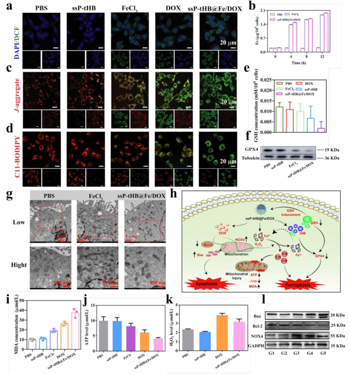
Antitumor mechanisms of ssP-tHB@Fe/DOX. (a) ROS levels (indicated by DCFH-DA) in 4T1 cells after various treatments using CLSM. (b) Analysis of intracellular Fe2+ content in 4T1 cells at different time points (0, 4, 8, and 12 h) after incubation with PBS, FeCl3, and ssP-tHB@Fe/DOX. (c) Confocal microscopic images of 4T1 cells stained with JC-1 probes to evaluate intracellular mitochondrial membrane potential. (d) CLSM images of C11-BODIPY-stained 4T1 cells after different treatments. Scale bar = 20 μm. (e) Analysis of the GSH/GSSG ratio in cells. (f) Western blot analysis of GPX4 expression. (g) Representative bio-TEM images of 4T1 cells. (h) The proposed schematic diagram of ssP-tHB@Fe/DOX-induced ferroptosis/apoptosis. (i, j, and k) Intracellular MDA levels (i), ATP content (j), and H2O2 concentration (k) of 4T1 cells after different treatments. (l) Western blot analysis of the expressions of Bax, Bcl-2, and NOX4. Note: PBS (G1), ssP-tHB (G2), FeCl3 (G3), DOX (G4), and ssP-tHB@Fe/DOX (G5)
Subsequently, treatments by different samples of 4T1 intracellular GSH levels were examined through GSH/GSSG assay kit. Figure 3e indicated that GSH levels significantly decreased compared to the PBS group following treatment with various samples, including DOX, FeCl3, ssP-tHB, and ssP-tHB@Fe/DOX. As anticipated, the consumption of endogenous GSH levels was optimal during treatment with ssP-tHB@Fe/DOX compared to other groups. This phenomenon occurred due to two main reasons: (i) the involvement of reduction reactions in groups containing Fe3+, and (ii) the consumption of nanoparticles through a sulfhydryl-dissulfhydryl exchange reaction with GSH.
Additionally, according to earlier reports, the consumption of GSH disrupts the equilibrium of intracellular redox system, leading to the emergence of cell oxidative stress and LPO [45]. We primarily monitored the generation of intracellular ROS utilizing 2’,7’-dichlorodihydrofluorescein diacetate (DCFH-DA) staining. As shown in Fig. 3a, the strong intensity of green fluorescence from DCF was displayed after incubation of ssP-tHB@Fe/DOX. Besides, other treated groups (FeCl3 and DOX) also showed low intensity of green signal compared to control group or ssP-tHB, demonstrating that the generation of ROS. To investigate the production of •OH via iron-mediated Fenton reaction within tumor cells, the fluorescence probe of HPF (indicating intracellular •OH levels) was employed [46]. As shown in Fig. S9, a notable increase in HPF green fluorescence was observed in the ssP-tHB@Fe/DOX group compared to PBS, ssP-tHB, FeCl3, and DOX groups. This analysis further confirmed that ssP-tHB@Fe/DOX-treated 4T1 cells induced the generation of abundant •OH through iron-mediated Fenton reaction, leading to oxidative damage of cell membranes and triggering ferroptosis.
Owing to the strong oxidative capacity of ROS in 4T1 cells, intracellular polyunsaturated fatty acids (PUFA) readily undergo oxidation, forming lipid peroxides (LPO), which leads to cell ferroptosis [45]. Therefore, we selected C11-BODIPY581/591 probe to detect LPO utilizing changes in the excitation emission wavelength in cells, to assess its ability to induce ferroptosis. Figure 3d demonstrated that red fluorescence decreased (reduction state) and green fluorescence increased (oxidized form) following ssP-tHB@Fe/DOX treatment, compared to other treatment groups, indicating the strongest LPO activity. Moreover, DOX-treated 4T1 cells demonstrated a similar level of LPO activity, aligning with the observed production of ROS. As expected, by malondialdehyde (MDA) concentration in 4T1 cells testing, similar results were observed (Fig. 3i). Since the Fe-mediated Fenton reaction depends on high levels of H2O2, we investigated the production of H2O2 in 4T1 cells by various samples. As already known, nicotinamide adenine dinucleotide phosphate oxidase 4 (NOX4), primarily derived from mitochondria, efficiently generates H2O2 by catalyzing the transfer of electrons from NADPH to oxygen [47, 48]. In early experiments, DOX not only induces cell apoptosis but also triggers oxidative stress by increasing the level of H2O2. A significant amount of H2O2 was produced during DOX treatment, contributing to the ROS generation in tumor cells induced by DOX [49]. In addition, DOX-containing nanoparticles also produced abundant H2O2, compared to blank carriers (ssP-tHB) (Fig. 3k). Therefore, in the tumor microenvironments, the circulatory amplification therapeutic strategies were formed, on the one hand, DOX induced chemotherapy, and on the other, DOX-assisted H2O2 production also aided in the amplification of ferroptosis.
The morphology of mitochondria in 4T1 cells treated with ssP-tHB@Fe/DOX was examined using bio-TEM. Compared to the PBS group, the ssP-tHB@Fe/DOX-treated mitochondria exhibited typical ferroptosis characteristics, including smaller size, increased membrane density, reduced or absent mitochondrial ridges, and disrupted outer mitochondrial membranes [50] (Fig. 3g). Figure 3c showed CLSM images of 4T1 cells after treatments of different samples and stained with 5,5′,6,6′-tetrachloro−1,1′,3,3′-tetraethylbenzimi-dazolylcarbocyanine iodide (JC−1) to monitor the intracellular mitochondrial membrane potential (MMP). The JC−1 showed red fluorescence after treatment with PBS or ssP-tHB, indicating a state of aggregation within mitochondria. However, the JC−1 showed increased green fluorescence after treatment with free FeCl3 or DOX alone, and the most intense green fluorescence when treated with ssP-tHB@Fe/DOX, suggesting the presence of monomers within mitochondria in 4T1 cells and indicating the strong •OH generation capability of ssP-tHB@Fe/DOX. This occurs because the •OH can harm the mitochondria, resulting in decreased MMP.
Extensive studies have identified glutathione peroxidase 4 (GPX4) as a classic indicator of ferroptosis [23, 24]. Abnormalities in intracellular mitochondrial activity, particularly in energy metabolism, were observed due to ssP-tHB@Fe/DOX-induced ferroptosis. The laboratory results indicated a significant decrease in ATP levels in 4T1 cells during ssP-tHB@Fe/DOX cultivation, compared to the control group. Other treatment groups (ssP-tHB, free FeCl3, and DOX alone) also showed the reduction of ATP levels (Fig. 3j). To better understand the mechanisms of ssP-tHB@Fe/DOX-induced cell death, specifically apoptosis and ferroptosis, we investigated the related pathways in pre-treated 4T1 cells. The results of the western blot (WB) assays visually demonstrated that ssP-tHB@Fe/DOX inhibits GPX4 expression in vitro (Fig. 3f). In the free FeCl3 treatment group, there was a lower reduction in GPX4 levels compared to the normal group, which mirrored the results of GSH consumption due to their positive correlation [39]. Additionally, the decrease in GXP4 expression in blank ssP-tHB also could be attributed to the consumption of GSH. Hence, the expression of levels of NADPH oxidase 4 (NOX4) in cells was investigated. As illustrated in Fig. 3l, S10c, DOX (G4) distinctly raised an enhancement in NOX4 level in comparison to the control group. Additionally, Additionally, treatment with ssP-tHB (G2) and iron ions (G3) showed minimal change. However, during the conversion of low-toxicity H2O2 into high-toxicity ·OH, ssP-tHB@Fe/DOX-treated mitochondria also experienced a certain degree of mitochondrial dysfunction, such as mitochondrial shrinkage and dissipation of membrane potential. Therefore, through WB analysis (Fig. S10c), we intriguingly observed that free DOX-induced NOX4 level was slightly higher than those induced by ssP-tHB@Fe/DOX. Subsequently, the impact of ssP-tHB@Fe/DOX on apoptotic pathways in 4T1 cells was further investigated. As illustrated in Fig. 3l, S10a, and S10b, G5 significantly mediated the increase in pro-apoptotic protein Bax and the decrease in anti-apoptotic protein Bcl−2. In other therapeutic groups, such as G2, G3, and G4, there was a decrease in anti-apoptotic Bcl−2 proteins and an increase in pro-apoptotic Bax proteins compared to the control group, indicating their ability to induce apoptosis to some extent.
Cellular internalization and cytotoxicity assays in vitro
Next, relevant cytotoxicity experiments in vitro were evaluated through CCK−8 assays (Fig. 4a). Blank ssP-tHB and tHB-treated 4T1 cells showed significant biocompatibility, even at a concentration of 640 µg/mL, with only a minor decrease in cell viability (Fig. S11). Subsequently, the normal L929 cells were used to investigate the tumor-specific characteristics of ssP-tHB@Fe/DOX and free DOX (Fig. 4b, S12). After 24 h of incubation, the CCK−8 measurement results showed that increasing concentrations of ssP-tHB@Fe/DOX showed minimal cytotoxicity, demonstrating significant biosafety for normal cells. However, due to the spectral anti-cancer effect of free DOX and its potent cytotoxicity, healthy L929 cells exhibited concentration-dependent cytotoxic effects (Fig. S12). Additionally, a dose-dependent cytotoxicity effect on 4T1 cells was exhibited in the presence of ssP-tHB@Fe/DOX, further indicating its tumor-specific therapeutic potential (Fig. 4b). As shown in Fig. 4c, trivalent iron ions (G3) also exhibited a degree of anti-tumor effect, with a cell survival rate of 72.7%, which may be attributed to iron-induced cell death through the Fenton reaction. In DOX-treated experiments (G4) at equal doses, ssP-tHB@Fe/DOX (G5, 125 µg/mL) demonstrated significantly greater cytotoxicity compared to free DOX.
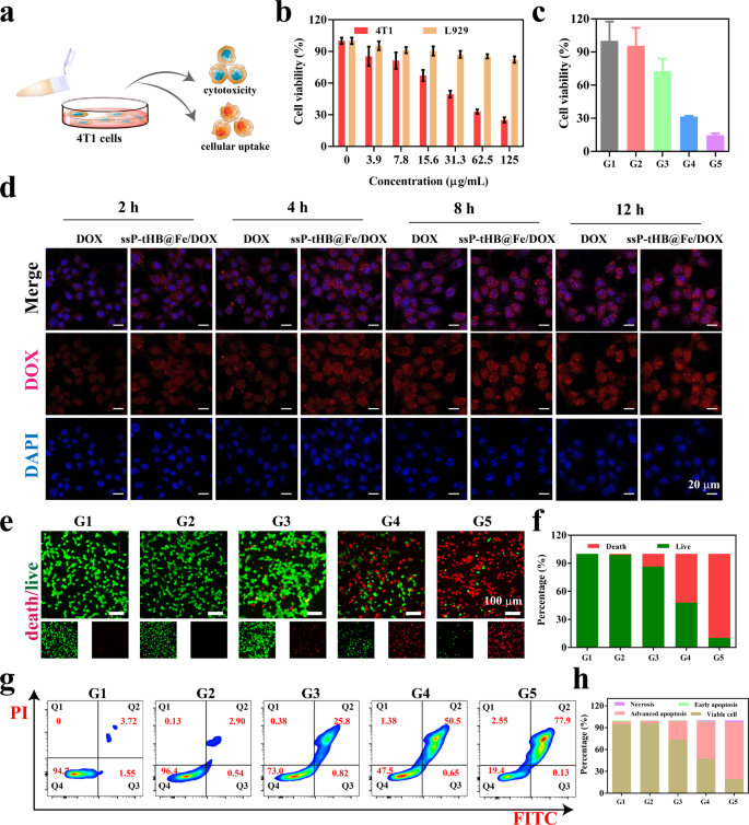
In vitro cytotoxicity related assessment. (a) The viability of 4T1 cells after different ssP-tHB concentrations treatment. (b) Cell viability of 4T1 cells and L919 cells treated with ssP-tHB@Fe/DOX for 24 h. (c) Relative viability of 4T1 cells in different samples. (d) Cellular uptake detected by CLSM. (e) CLSM images of Calcein AM/PI-stained 4T1 cells after different treatments. (f) Quantification of live and dead cells after treatment. (g) 4T1 cell apoptosis assessed through annexin V-FITC and PI staining, analyzed by FCM. (h) Histogram showing quantification of different apoptotic forms. Note: PBS (G1), ssP-tHB (G2), FeCl3 (G3), DOX (G4), and ssP-tHB@Fe/DOX (G5)
Cellular uptake behavior is considered a crucial indicator for assessing the anti-tumor efficacy of “nano-vehicles” [51]. Therefore, as free DOX or ssP-tHB@Fe/DOX-cultured time increased, the intensity of red fluorescence signals became stronger by CLSM images (Fig. 4a and d). Simultaneously, for free DOX, 4T1 cells showed a time-dependent increase in fluorescence. Importantly, as the culture time increases, red fluorescence (DOX) overlapped with blue fluorescence (nucleus), indicating that DOX released from ssP-tHB@Fe/DOX had moved from the cytoplasm to the nucleus for chemotherapy. The effect of phagocytosis of ssP-tHB@Fe/DOX was more pronounced compared to free DOX, likely due to the enhanced cellular uptake facilitated by nanoparticles.
Calcein-AM (green fluorescence for live cells) and propidium iodide (PI, red fluorescence for dead cells) were used together to differentiate between live and dead 4T1 cancer cells. As shown in Fig. 4e and f, after processing with blank ssP-tHB (G2), nearly all green fluorescence signals were observed, indicating its non-toxic nature. Free FeCl3 (G3) and DOX (G4) were found to kill cells to some extent, but their therapeutic effect was limited compared to the ssP-tHB@Fe/DOX. The results aligned with the conclusions of the CCK−8 assay.
Given that the chemotherapy drug DOX effectively induces apoptosis in tumor cells [42, 47], we also examined the apoptosis of different samples on 4T1 cells using flow cytometry (Fig. 4g and h). Compared to the control group (G1) or blank NPs (G2), the proportion of apoptotic cells significantly increased in the FeCl3-treated group (G3). Furthermore, free DOX (G4) showed strong cell apoptosis, with the apoptosis rate of 50.5%. Of note, the cell apoptosis rate rose to 77.9% after treatment with ssP-tHB@Fe/DOX (G5).
Hemolysis and acute toxicity test in vivo
Inspired by the brilliant performance of ssP-tHB@Fe/DOX against 4T1 cells in vitro, its anti-tumor efficacy was further assessed in vivo. To investigate the biosafety of ssP-tHB@Fe/DOX, we performed hemolysis tests and acute toxicity measurements in vivo. The hemolysis rates were low, at less than 10%, compared to the control group. At a concentration of 200 µg/mL, the ssP-tHB@Fe/DOX caused a hemolysis rate of only 8.78%, indicating that the erythrocyte structure remained normal (Fig. S13). As a result, ssP-tHB@Fe/DOX, with its tumor-specific therapy, demonstrated significant blood compatibility and can be further utilized in in vivo bio-related assessments. Subsequently, female Balb/c mice were randomly divided into two groups (n = 6), and then injected intravenously with PBS (control group), ssP-tHB@Fe/DOX (DOX: 6 mg/kg, 100 µL), respectively. After 8 and 14 days of treatment, all mice were euthanized, and their major organs (including the heart, liver, spleen, lung, and kidney) were removed for hematoxylin and eosin (H&E) staining (Fig. S14), and blood was also collected from their eyeballs for biochemical analysis (Tables S1, S2) [52]. The H&E analysis revealed no significant organ damage or inflammation in the ssP-tHB@Fe/DOX-treated mice. Furthermore, ssP-tHB@Fe/DOX injection had no effect on liver and kidney functions or blood parameters. In summary, the GSH/pH dual-responsive ssP-tHB@Fe/DOX showed significant potential for biological applications.
RNA-seq analysis of ssP-tHB@Fe/DOX
RNA sequencing (RNA-seq) analysis was conducted to evaluate the therapeutic mechanism of ssP-tHB@Fe/DOX in 4T1 cells. As shown in Fig. 5a, the heat map of mRNA reveals changes in the cell transcriptome induced by treatment with ssP-tHB@Fe/DOX. Moreover, principal component analysis (PCA) revealed a significant clustering relationship between the ssP-tHB@Fe/DOX treatment group and the PBS control group (Fig. 5b). We then conducted bioinformatics analyses to compare the differences (p-value 2 FC| > 1) (Fig. 5c). The results of the collection found that the threshold of differentially expressed genes (DEGs) under ssP-tHB@Fe/DOX-mediated 4T1 cells was 3373, of which 1873 gene up-regulated, 2101 gene down-regulated. Then, the key pathways involved in the therapeutic effect of ssP-tHB@Fe/DOX were analyzed through enrichment analysis. Gene Ontology (GO) enrichment analysis (Fig. S15) and pathway enrichment analysis using the Kyoto Encyclopedia of Genes and Genomes (KEGG) database (Fig. 5g) revealed significant enrichment in p53 signaling pathways, apoptosis-related processes, and amino acid metabolism. Therefore, the obtained results showed that treatment with ssP-tHB@Fe/DOX in 4T1 cells could partially exert its anti-cancer effect by activating p53 signaling to induce cellular apoptosis and by modulating amino acid metabolism to induce ferroptosis.
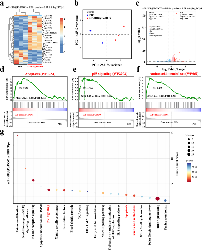
(a) Heat map of mRNAs related to treatment progression. (b) PCA based on bulk RNA-seq results of the PBS and ssP-tHB@Fe/DOX. (c) Volcano map of the transcriptomic profiles. (d, c, and f) GSEA of apoptosis (WP1254) (d), p53 signaling (WP2902) (e), and amino acid metabolism (WP662) (f) signaling pathway after ssP-tHB@Fe/DOX therapy. (g) Bubble diagram of differentially expressed genes enriched in KEGG
In addition, to better illustrate the transcriptome changes induced by ssP-tHB@Fe/DOX, we conducted gene set enrichment analysis (GSEA) on the RNA-seq data. As expected, the three significantly enriched signature pathways, such as apoptosis (WP1254) (Fig. 5d), p53 signaling (WP2902) (Fig. 5e), and amino acid metabolism (WP662) (Fig. 5f), were displayed. More encouragingly, gene regulators involved in cell apoptosis and ferroptosis mediated by ssP-tHB@Fe/DOX, such as drivers, suppressors, and markers, were identified (Fig. S16, S17, and S18). Ultimately, we could speculate that breast cancer could be effectively treated through ssP-tHB@Fe/DOX by inducing both apoptosis and ferroptosis.
Therapeutic efficiency against 4T1 tumors and biosafety in vivo
To evaluate the therapeutic efficacy of the cyclic magnification ferroptosis and DOX-induced chemotherapy in ssP-tHB@Fe/DOX, the treatment’s effect on a subcutaneous 4T1 tumor model was assessed in vivo. Firstly, 4T1 tumor-bearing mice were successfully established (approximately tumor volume: 80 mm3) and were randomly assigned five groups, PBS group (G1), ssP-tHB (G2), FeCl3 (G3), DOX (G4), and ssP-tHB@Fe/DOX (G5, DOX: 6 mg/kg), respectively. Afterwards, we administered the diverse samples with an equal volume via intravenous injections every two days (five times in total) for a total of 17 days and recorded the tumor volume changes and body weight fluctuations, as visually depicted in Fig. 6a. Throughout the treatment period, 4T1 tumor-bearing mice in all groups showed venial upward trend in weight (Fig. 6b), suggesting that the therapeutic samples did not interfere with normal growth in mice. The tumor growth curves showed that, unlike the PBS (G1) and ssP-tHB (G2) groups, which exhibited rapid tumor growth, the other therapeutic groups (G3, FeCl3; G4, DOX; and G5, ssP-tHB@Fe/DOX) demonstrated significant tumor volume reduction. Among these, G5 (ssP-tHB@Fe/DOX) was the most effective, aligning with results from in vitro cell experiments (Fig. 6f and g). Actually, this supports the results that ferroptosis-chemotherapeutic combination therapy is superior to mono-therapy. This observation was further supported by digital photographs of the tumors taken 17 days after treatment with different samples, as shown in Fig. 6c. At 14 days post-treatment, as shown in Fig. 6d and e, G5 demonstrated the most significant tumor inhibition due to the combined effects of ssP-tHB@Fe/DOX-induced efficient ferroptosis and DOX-mediated apoptosis.
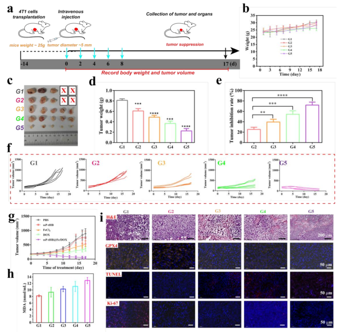
Anti-tumor treatments with ssP-tHB@Fe/DOX in 4T1 tumor model. (a) Schematic illustration of the in vivo antitumor treatment experiment. (b) Body-weight changes of mice (n = 5). (c) Representative tumor images of 4T1 tumor-bearing mice receiving various treatments. (d) Weights of tumors harvested from 4T1 tumor-bearing mice in different groups. (e) Tumor growth inhibition rate for each treatment group after 17 days of treatment. (f) Spaghetti curves showing tumor growth in mice treated differently. (j) Tumor growth curves (n = 5) for tumor-bearing mice injected intravenously with G1, G2, G3, G4, and G5, respectively. (h) MDA content in serum of mice from each treatment group post-treatment. (i) Histological images of H&E, and TUNEL, antigen Ki-67, GPX4 immunofluorescence-stained tumor slices from the 4T1 tumor-bearing mice at day 17. PBS (G1), ssP-tHB (G2), FeCl3 (G3), DOX (G4), and ssP-tHB@Fe/DOX (G5), **p p p
Subsequently, for specific analysis of pathology, the collected tumors were stained via hematoxylin and eosin (H&E), TdT-mediated dUTP Nick-End Labeling (TUNEL), Ki−67, and GPX4 (Fig. 6i). As anticipated, H&E and TUNEL imaging revealed that the ssP-tHB@Fe/DOX group (G5) exhibited a higher level of apoptosis compared to the other groups. The cell proliferation ability of ssP-tHB@Fe/DOX exhibited a significant decrease, as indicated by Ki−67 protein staining, compared to other treatment groups. These results showed that ssP-tHB@Fe/DOX with multi-mode combination exhibits significant anti-tumor effects. Next, we assessed the expression of GPX4 in different samples-treated tumors. Comparatively, the expression of GPX4 in tumors was receded by ssP-tHB@Fe/DOX (G5) treatment, proving its ability to enhance ferroptosis (Fig. 6i). MDA is natural byproducts of LPO. We measured the MDA expression levels in the serum of each group after treatment. As presented in Fig. 6h, compared to the PBS group, the expression of MDA was the highest in the G5, which again indicates that ssP-tHB@Fe/DOX could enhance LPO and lead to ferroptosis.
The biosafety of mice with 4T1 tumors was also a crucial factor in the treatment process. Therefore, the functions of liver (indicators: alkaline phosphatase (ALP), alanine transaminase (ALT), aspartate transaminase (AST)) and kidney (indicators: creatinine (CREA), urea (UREA)) were analyzed, as well as routine bloods (indicators: platelet hematocrit (PCT), white blood cell (WBC) and red blood cell (RBC)) of mice were detected [52]. As shown in Fig. S19, the levels of ALP, ALT, and AST were significantly elevated in DOX alone-treated mice (G4), indicating the induction of hepatotoxicity. No significant changes were observed in the levels of ALT, ALP, AST, UREA, CREA, PCT, WBC, and RBC in the G2, G3, and G5 treatment groups. These data indicated no marked toxic effects at the current dose of ssP-tHB@Fe/DOX. Correspondingly, H&E staining results indicated significant liver damage exclusively in the DOX treatment group, whereas no notable injuries were observed in the livers, kidneys, or hearts of mice treated with ssP-tHB@Fe/DOX (Fig. S20). These results indicated that ssP-tHB@Fe/DOX had an excellent biocompatibility.


