Morphological and structural analysis of Pd@CeO2
Pd@CeO2 nanozymes were synthesized via a multi-step process depicted in Fig. 1a. Uniform Pd nanocubes (~ 18 nm) were initially formed by reducing Na2PdCl4 using ascorbic acid, polyvinylpyrrolidone, and potassium bromide, ensuring monodispersed and precisely sized nanoparticles (Fig. 1b–c). Subsequently, cerium nitrate was introduced in the presence of L-Arginine as a capping agent, facilitating CeO2 nanoparticles’ self-assembly onto the Pd nanocube surfaces. This process resulted in a distinct core-shell Pd@CeO2 structure, confirmed by TEM imaging showing complete and uniform encapsulation of Pd cores by CeO2 shells (Fig. 1d–e). Energy-dispersive X-ray spectroscopy (EDS) elemental mapping (Fig. 1f) demonstrated the spatial distribution of Pd, Ce, and O, further confirming successful core-shell formation. The crystallinity of Pd and Pd@CeO2 phases was validated through X-ray diffraction (XRD), which exhibited characteristic peaks corresponding to both materials (Figure S1). Dynamic light scattering (DLS) measurements confirmed that Pd@CeO2 nanozymes exhibited a mean particle diameter of 103.9 ± 0.56 nm (Fig. 1g), whereas the zeta potential measurement indicated a surface charge of −19.5 ± 0.47 mV (Fig. 1h).
To elucidate the electron-injection mechanism, X-ray photoelectron spectroscopy (XPS) was employed. Analysis of the Ce 3 d spectra revealed a marked increase in the Ce3+/Ce4+ ratio in Pd@CeO2 (34.23%/65.77%) compared with pristine CeO2 (21.29%/78.71%). Furthermore, peak fitting of the Ce 3d spectra revealed that Pd@CeO2 exhibits overall lower binding energies than CeO2 (Fig. 1i). In particular, the enhanced intensity of Ce3+ peaks at these shifted positions indicates electron enrichment at the CeO2 interface, arising from interfacial electron transfer from Pd to CeO2, which provides robust ROS-scavenging performance. Stability assessments over 7 days revealed minimal particle aggregation or charge alterations, underscoring the nanoparticle’s suitability for physiological applications (Figure S2).
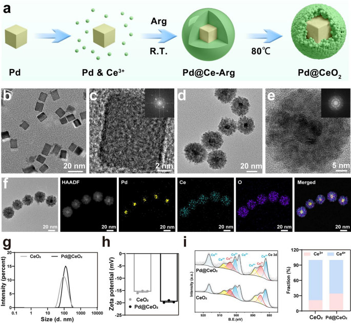
Synthesis and characterization of Pd@CeO2 nanozymes. (a) Schematic illustration of the Pd@CeO2 nanozymes preparation process. (b) TEM images of Pd nanocubes. (c) Electron diffraction (SAED) pattern of Pd nanocubes. (d) TEM images of Pd@CeO2 nanozymes. (e) SAED pattern of Pd@CeO2 nanozymes. (f) Element mapping of Pd, Ce, O, and the merged image of these three elements. (g–h) Particle size distribution and zeta potential of Pd@CeO2 nanozymes. (i) XPS of Pd@CeO2 nanozymes
Enzyme-mimicking activities of Pd@CeO2 and mechanistic insights from DFT calculations
ROS including hydroxyl radicals (·OH), superoxide anions (·O2⁻), and hydrogen peroxide (H2O2), are pivotal in numerous biological and pathological processes [31, 32]. We first evaluated the ·OH scavenging capability of Pd@CeO2 using electron spin resonance (ESR) spectroscopy. ESR spectra (Fig. 2a) displayed a distinct 1:2:2:1 quartet signal characteristic of DMPO-·OH adducts generated through the Fenton reaction. Compared to controls, Pd@CeO2 exhibited significantly reduced ESR signal intensity, demonstrating superior ·OH scavenging due to the enhanced catalytic effect of Pd@CeO2. Subsequently, the SOD-like activity of Pd@CeO2 was investigated. ESR spectra of ·O2⁻ (Fig. 2c) revealed a pronounced reduction in the typical 1:1:1:1 quartet upon treatment with Pd@CeO2, confirming efficient superoxide neutralization. Quantitative analysis using the nitro blue tetrazolium (NBT) assay (Fig. 2d–e) further validated this enhanced performance, as Pd@CeO2 exhibited the lowest absorbance at 560 nm, reflecting the highest inhibition rate (48.72%) compared with CeO2 (24.81%) and Pd + CeO2 mixtures (37.18%). CAT-like activity, essential for H2O2 detoxification, was evaluated using a colorimetric assay (Fig. 2g–h). Pd@CeO2 showed the lowest absorbance at 405 nm and a markedly higher H2O2 inhibition rate (77.1%) compared to CeO2 (40.8%) and Pd + CeO2 (44.7%). Additionally, dissolved oxygen measurements revealed rapid catalytic decomposition of H2O2 to O2 upon Pd@CeO2 treatment, increasing O2 concentrations from 7.5 µg/mL to 28.3 µg/mL within 20 min—significantly outperforming CeO2 nanoparticles (21.2 µg/mL) (Fig. 2i). These results demonstrate that Pd@CeO2 exhibits comprehensive ROS scavenging capacity, integrating ·OH, ·O2⁻, and H2O2 elimination through synergistic multi-enzyme–like activities.
To elucidate the mechanism underlying the exceptional catalytic performance of the CeO2 (111) slab decorated with Pd clusters, density functional theory (DFT) calculations were performed. A Pd@CeO2 model was constructed and compared with the pristine CeO2 slab (Fig. 3a). Owing to the pronounced empty-band distribution in CeO2, the position of the d-band center relative to the Fermi level was carefully considered. For the CeO2 (111) slab, the d-band center is located at 3.13 eV (Fig. 3b). Upon adsorption of a Pd cluster, the d-band center shifts downward by 0.38 eV to 2.75 eV (Fig. 3c), indicating a weakened adsorption strength of oxygen intermediates at the surface Ce sites. These results demonstrate that the incorporation of Pd clusters induces pronounced electronic interactions within the catalyst, resulting in a downward shift of the d-band center. This adjustment optimizes the adsorption of essential intermediates and thereby enhances catalytic efficiency, as illustrated in Fig. 3d. The charge density difference analysis further confirms the electronic modulation induced by the Pd clusters. As shown in Fig. 3e, adsorption of a Pd cluster results in a charge transfer of approximately 1.15 electrons from the cluster to the CeO2 substrate, as determined by Bader charge analysis. This electron donation increases the electron density on the substrate, strengthening electronic interactions on CeO2. This observation is consistent with the projected density of states (PDOS) analysis, which reveals the downward shift of the d-band center. Furthermore, the adsorption energies of oxygen intermediates on the Pd@CeO2 surface were systematically evaluated. The reaction energy barriers of each elementary step were also calculated, revealing a clear relationship between the overlap of transition-metal d orbitals and the adsorption of oxygen intermediates on the catalyst surface (Fig. 3f). For both CeO2 and Pd@CeO2 systems, we found that the rate-determining step (RDS) for H2O2 decomposition producing ·OH radicals (i.e., the peroxidase-like reaction) is associated with higher energy barriers compared to H2O2 decomposition producing O2 (i.e., the CAT-like reaction), with values of 1.93 eV vs. 1.14 eV and 1.42 eV vs. 1.08 eV, respectively. This result suggests that the CAT-like pathway predominates in the overall reaction. Notably, the adsorption of Pd clusters significantly reduces the RDS. Specifically, the formation of *O is identified as the RDS for Pd@CeO2, with a calculated energy barrier of 1.08 eV, which is lower than the 1.14 eV barrier observed in the pristine CeO2 system. This reduction can be attributed to the enhanced electron density at the Ce sites induced by Pd cluster adsorption, which is consistent with the Bader charge analysis, and facilitates H2O2 reduction and consequently promotes O2 evolution, thereby conferring superior CAT-like activity. These findings provide fundamental insights into how Pd clusters regulate the electronic structure of CeO2, thereby improving catalytic activity.
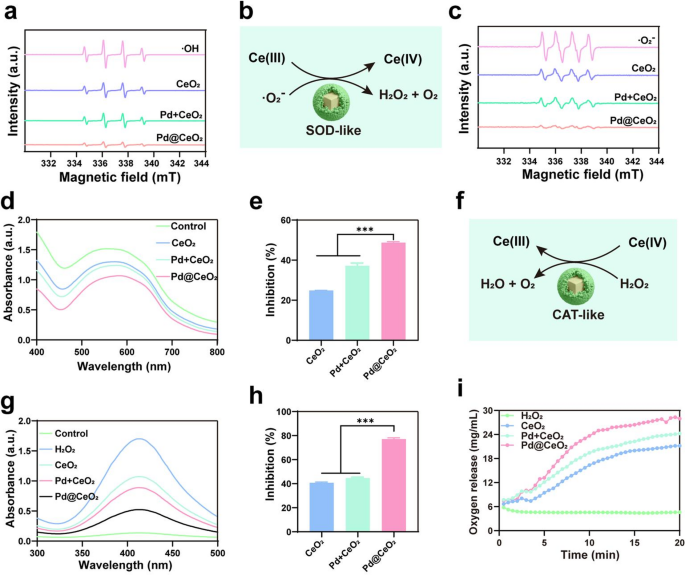
ROS elimination by Pd@CeO2 nanozymes. (a) ESR spectra of ·OH radicals trapped by DMPO in different samples (positive group, CeO2, Pd + CeO2 and Pd@CeO2). (b) Schematic representation of the SOD-like catalytic mechanism of Pd@CeO2. (c) ESR spectra of ·O2− scavenging activity for CeO2, Pd + CeO2, and Pd@CeO2. (d) NBT assay showing the reduction of ·O2− by Pd@CeO2 through decreased absorbance at 560 nm. (e) Inhibition rates of ·O2− by different formulations. (f) Schematic of the CAT-like catalytic mechanism of Pd@CeO2. (g) CAT-like activity of Pd@CeO2 was evaluated using a catalase assay kit, where the decrease in absorbance at 405 nm reflects H2O2 decomposition. (h) Inhibition rates of H2O2 decomposition by CeO2, Pd + CeO2, and Pd@CeO2. (i) Oxygen generation during H2O2 decomposition by CeO2 NPs, Pd + CeO2 NPs, and Pd@CeO2
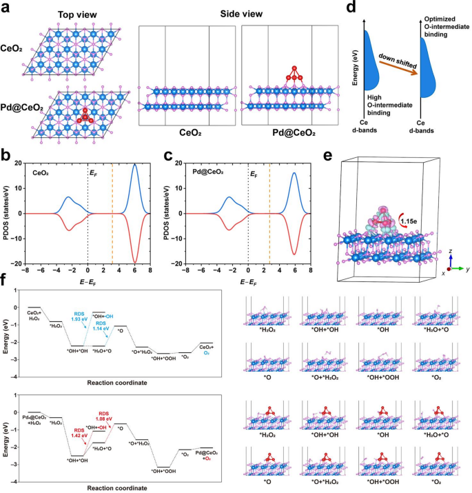
DFT-based mechanistic understanding of Pd@CeO2 catalysis. (a) Models of the CeO2 (111) slab (denoted as CeO2) and the CeO2 (111) slab with an adsorbed Pd cluster (denoted as Pd@CeO2). Spheres in teal, red and grey represent cerium, oxygen and palladium, respectively. (b–c) PDOS plots of the (b) CeO2 and (c) Pd@CeO2. (d) Schematic illustration showing that the d-band center shift of CeO2 induced by Pd4 modulates the binding ability of O-containing intermediates. (e) Charge density difference distribution of the Pd@CeO2, and insert is Bader charge analysis of Pd4 on CeO2 (111) slab. The pink and cyan volumes represent electron depletion and accumulation, respectively. Spheres in teal, red and grey represent cerium, oxygen and palladium, respectively. (f) Energy profiles and the corresponding configurations for the formation of surface peroxo species on the CeO2 (111) slab, either pristine (denoted as CeO2) or decorated with a Pd cluster (denoted as Pd@CeO2). The rate-determining step (RDS) is highlighted in the plots. Spheres in blue, white, pink and red represent cerium, hydrogen, oxygen and palladium, respectively
In vitro ROS-scavenging capability of Pd@CeO2 nanozymes
Building on the robust enzyme-mimetic catalytic activities of Pd@CeO2, we evaluated their cellular uptake, biocompatibility, and cytoprotective performance against oxidative stress. The internalization of Pd@CeO2 nanozymes was monitored in HUVEC and RAW 264.7 cells using FITC labeling. Fluorescence microscopy and flow cytometry consistently demonstrated efficient, time-dependent cellular uptake of Pd@CeO2, with uptake efficiency reaching over 95% within 12 h and approaching complete internalization by 24 h (Fig. 4a–b). Biocompatibility assessment using CCK-8 assays revealed negligible cytotoxicity of Pd@CeO2 at concentrations ≤ 50 µg/mL in both HUVEC and RAW 264.7 cells (Fig. 4c–d). Longitudinal analysis over a three-day period confirmed the absence of adverse effects on cell proliferation, highlighting the suitability of Pd@CeO2 for biomedical applications (Figure S3). Next, we evaluated the protective role of Pd@CeO2 against H2O2-induced oxidative injury. Pretreatment with Pd@CeO2 markedly enhanced HUVEC viability (91.66%), outperforming CeO2 (72.72%) and Pd + CeO2 mixtures (79.79%) (Fig. 4e). Consistently, intracellular ROS levels, visualized by DCFH-DA fluorescence, were substantially diminished in the Pd@CeO2 group, underscoring its superior scavenging efficacy relative to the controls (Fig. 4f and i and Figure S4–S5). Calcein-AM/PI staining further confirmed enhanced cell survival and reduced cell death following Pd@CeO2 treatment under oxidative stress conditions (Fig. 4g and j). Flow cytometry analyses using Annexin V-FITC/PI staining demonstrated a substantially lower apoptosis rate in Pd@CeO2-treated cells, highlighting robust anti-apoptotic effects (Fig. 4h and k). Collectively, the findings establish Pd@CeO2 as a safe and efficient nanozymes with superior antioxidant and anti-apoptotic properties, highlighting its promise for biomedical applications in oxidative stress–driven pathologies.
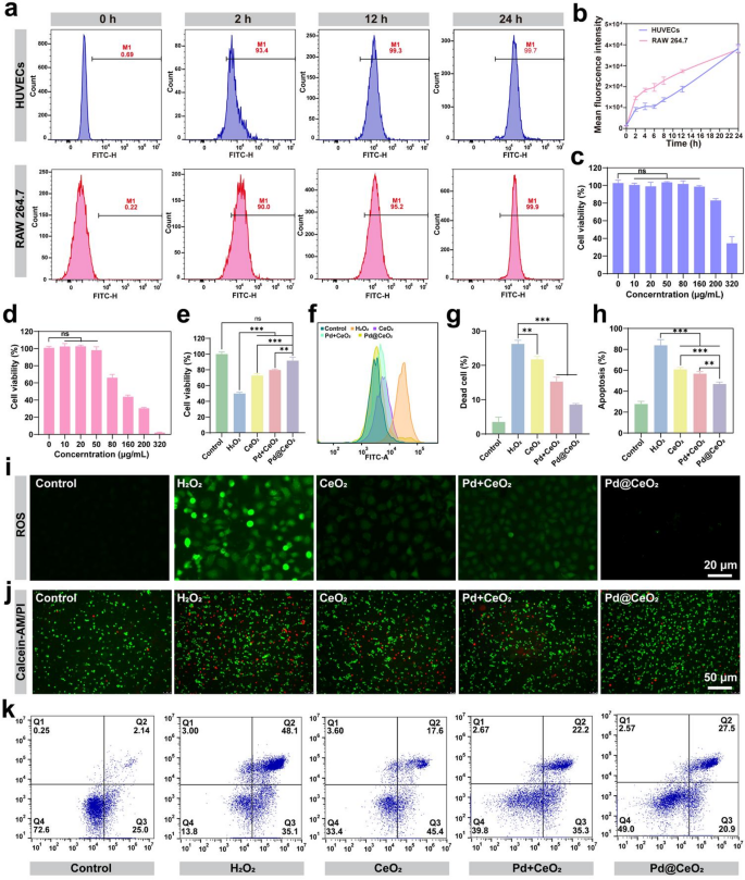
Biocompatibility and protective effects of Pd@CeO2 against oxidative damage. (a) Flow cytometry analysis of FITC-labeled Pd@CeO2 uptake by HUVECs and RAW 264.7 macrophages at different time points (0, 2, 12, and 24 h). (b) quantification of flow cytometry results. (c) Cytotoxicity of Pd@CeO2 on HUVECs at various concentrations (0, 10, 20, 50, 80, 160, 200, and 320 µg/mL). (d) Cytotoxicity of Pd@CeO2 on RAW 264.7 macrophages at various concentrations (0, 10, 20, 50, 80, 160, 200, and 320 µg/mL). (e) Cell viability of CeO2, Pd + CeO2, and Pd@CeO2 (50 µg/mL) against H2O2-induced oxidative damage. (f) Flow cytometric analysis of intracellular ROS levels in oxidatively stressed HUVEC cells. (g) Statistical analysis of cell mortality in injured cells. (h) Statistical analysis of apoptosis rate in each group. (i) DCFH-DA fluorescence microscopy images showing ROS levels in each group. Scale bars: 20 μm. (j) Calcein-AM/PI fluorescence staining of HUVECs treated with H2O2 and or different formulations. Scale bars: 50 μm. (k) Flow cytometry analysis of apoptosis rates after different treatments
Molecular mechanisms of Pd@CeO2 in suppressing apoptosis and inflammation
To further elucidate the protective mechanisms conferred by Pd@CeO2 nanozymes against oxidative stress-induced injury, we evaluated their influence on apoptosis-related pathways. Apoptosis, prominently regulated by proteins including Bcl-2 (anti-apoptotic), Bax (pro-apoptotic), and cleaved Caspase-3 (executioner of apoptosis), is central to I/R injury pathology [33, 34]. Western blot analysis (Fig. 5a–d) demonstrated that Pd@CeO2 significantly elevated Bcl-2 expression while concurrently suppressing Bax and cleaved Caspase-3 levels compared to CeO2 and Pd + CeO2. These findings indicate a robust inhibition of apoptosis via modulation of the Bax/Bcl-2/caspase-3 pathway, underscoring the potent cytoprotective capabilities of Pd@CeO2.
Inflammation represents another pivotal aspect of I/R injury, which is further aggravated by excessive ROS generation [35, 36]. Employing an LPS-stimulated RAW 264.7 macrophage inflammation model, ELISA assays revealed that Pd@CeO2 markedly attenuated secretion of key pro-inflammatory cytokines TNF-α, IL-1β, and IL-6 compared to the LPS-treated control groups (Fig. 5e–g). This highlights the substantial anti-inflammatory efficacy of Pd@CeO2 in cellular models. To uncover the molecular basis underlying these anti-inflammatory effects, quantitative PCR and Western blot analyses assessed the expression of inflammation-related factors NF-κB and PPAR-γ. The LPS-driven increase in NF-κB mRNA expression was substantially suppressed by Pd@CeO2, highlighting its robust capacity to modulate this pivotal pro-inflammatory transcription factor (Fig. 5h). Conversely, Pd@CeO2 significantly upregulated PPAR-γ mRNA, supporting its role in resolving inflammation (Fig. 5i). Protein-level analyses further confirmed these results, demonstrating reduced phosphorylation of NF-κB p65 and increased PPAR-γ protein expression in Pd@CeO2-treated groups compared to controls (Fig. 5j–l). These results suggest that Pd@CeO2 modulate inflammatory responses by downregulating the NF-κB pathway and upregulating the anti-inflammatory PPAR-γ pathway.
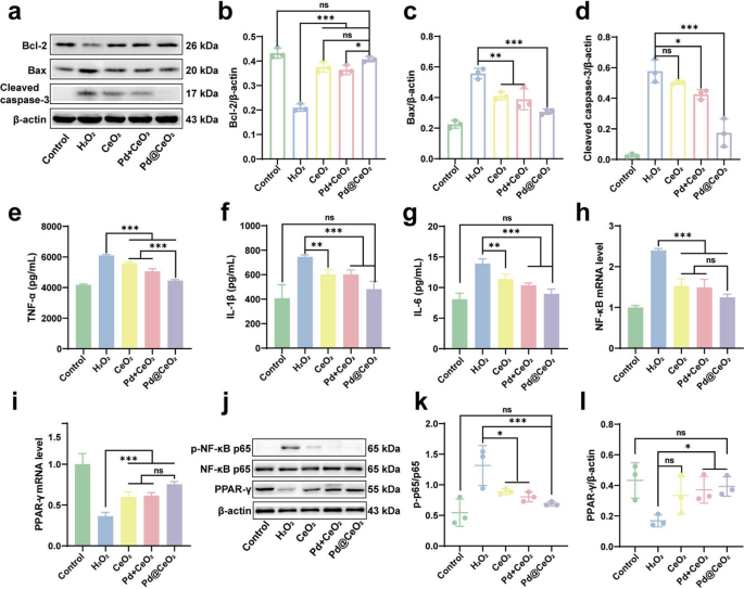
Anti-apoptotic and anti-inflammatory mechanisms of Pd@CeO2. (a) Western blot analysis of Bcl-2, Bax, and cleaved caspase-3 protein expression levels. Quantification of (b) Bcl-2, (c) Bax, and (d) caspase-3 protein expression levels. ELISA results showing (e) TNF-α, (f) IL-1β, and (g) IL-6 secretion levels after treated with CeO2, Pd + CeO2, or Pd@CeO2. Quantification of (h) NF-κB and (i) PPAR-γ mRNA expression levels by qPCR. (j) Western blot analysis of p-NF-κB p65, NF-κB p65, and PPAR-γ protein expression. Quantification of (k) p-p65 and (l) PPAR-γ protein expression ratio
Restoration of angiogenic capacity by Pd@CeO2 in oxidative environments
Building upon the demonstrated anti-apoptotic and anti-inflammatory properties of Pd@CeO2, we further investigated its angiogenic capacity under oxidative stress, a critical factor in tissue repair and flap survival. Angiogenesis is notably impaired by I/R injury, thus emphasizing its importance in recovery [37, 38]. qPCR analysis revealed that Pd@CeO2 treatment markedly upregulated VEGFA mRNA, a key driver of angiogenesis, in H2O2-stimulated HUVECs compared with controls, CeO2, and Pd + CeO2 (Fig. 6a). Western blot analyses corroborated this, revealing substantially higher VEGFA protein levels in Pd@CeO2-treated cells relative to H2O2-exposed cells (Fig. 6b–c). These results highlight the strong capacity of Pd@CeO2 to reestablish angiogenic signaling pathways disrupted by oxidative stress. We next evaluated endothelial cell migration using scratch assays. H2O2 exposure markedly impaired migratory capacity, whereas treatment with CeO2, Pd + CeO2, and especially Pd@CeO2 significantly promoted wound closure (Fig. 6d). Experiments further confirmed that Pd@CeO2 achieved the most pronounced enhancement of migration, surpassing all other treatments (Fig. 6f). Finally, we assessed angiogenic functionality via endothelial tube formation assays. H2O2-induced oxidative stress severely disrupted tube formation, evident by reduced branching and network complexity. Notably, pretreatment with Pd@CeO2 effectively restored endothelial tube formation, significantly enhancing total branching length compared to CeO2 and Pd + CeO2 treatments ((Fig. 6e and g). These collective results position Pd@CeO2 as a highly effective nanozyme, capable of significantly promoting angiogenesis under oxidative stress, thereby highlighting its substantial therapeutic potential in ischemic tissue repair.
In vitro investigations revealed that Pd@CeO2 orchestrates multifaceted cytoprotective mechanisms by reducing oxidative stress, inhibiting apoptosis through the Bcl-2/Bax/caspase axis, and dampening inflammatory cascades mediated by NF-κB and PPAR-γ (Fig. 6h), thereby establishing a coherent mechanistic basis for its therapeutic potential.
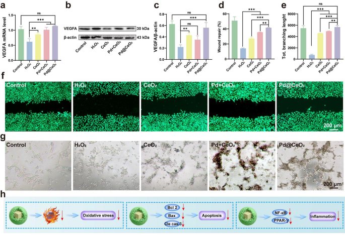
Enhancement of angiogenesis in oxidative stress-damaged HUVECs by Pd@CeO2. (a) qPCR analysis of VEGFA mRNA expression following treatment with CeO2, Pd + CeO2, or Pd@CeO2. (b) Western blot assessment of VEGFA protein expression. (c) Quantification of VEGFA protein expression level. (d) Statistical analysis of wound repair rate of each group. (e) Statistical evaluation of the total dendritic branching length across experimental groups. (f) Representative images of the scratch assay in HUVECs under H2O2-induced oxidative stress. Scale bar: 200 μm. (g) Representative images of tube formation assays in HUVECs exposed to H2O2. Scale bar: 200 μm. (h) Diagrammatic representation of Pd@CeO2-mediated mechanisms in attenuating oxidative stress, inhibiting apoptosis, and suppressing inflammation
Therapeutic efficacy of Pd@CeO2 in a rat skin flap ischemia–reperfusion injury model
To preliminarily assess the in vivo biosafety of Pd@CeO2 nanozymes, we examined their biocompatibility through hemocompatibility and histological studies. Hemolysis assays revealed an exceptionally low hemolysis rate (S1-S2). Histological examination of major organs (heart, liver, spleen, lung, and kidney) by H&E staining at 7 and 14 days revealed no detectable pathological alterations, thereby reinforcing the excellent biosafety profile of Pd@CeO2 (Figure S7).
Subsequently, therapeutic efficacy was evaluated using a rat dorsal skin flap I/R injury model. After 6 h of ischemia followed by reperfusion, extensive flap necrosis was observed in the untreated controls, whereas Pd@CeO2 treatment markedly preserved tissue integrity and minimized necrotic damage (Fig. 7a-b). Real-time microcirculation assessed via laser speckle imaging revealed significantly enhanced blood perfusion in the Pd@CeO2 group, which achieved the highest flap survival rate (85.42%) compared to CeO2 (60.10%), Pd + CeO2 (77.14%), and untreated groups (55.76%) (Fig. 7b). H&E staining further demonstrated significant preservation of tissue architecture in Pd@CeO2-treated flaps, characterized by intact epidermal layers, well-maintained hair follicles, and normal dermal collagen deposition, contrasting sharply with severe epidermal thinning in control groups (Fig. 7c). Given the promising in vitro angiogenic effects of Pd@CeO2, we subsequently evaluated its pro-angiogenic capabilities within an in vivo I/R skin flap model. Immunohistochemical staining for the endothelial marker CD31 was performed to assess neovascularization within flap tissues. Results indicated compromised angiogenesis in the I/R group, exhibiting microvascular densities of approximately 4.0 vessels per field. In contrast, groups treated with Pd + CeO2 and particularly Pd@CeO2 displayed considerably enhanced neovascularization, with microvessel densities of 13.5 and 16.0, respectively (Fig. 7d). These findings underscore the superior efficacy of Pd@CeO2 in promoting angiogenesis under I/R injury conditions.
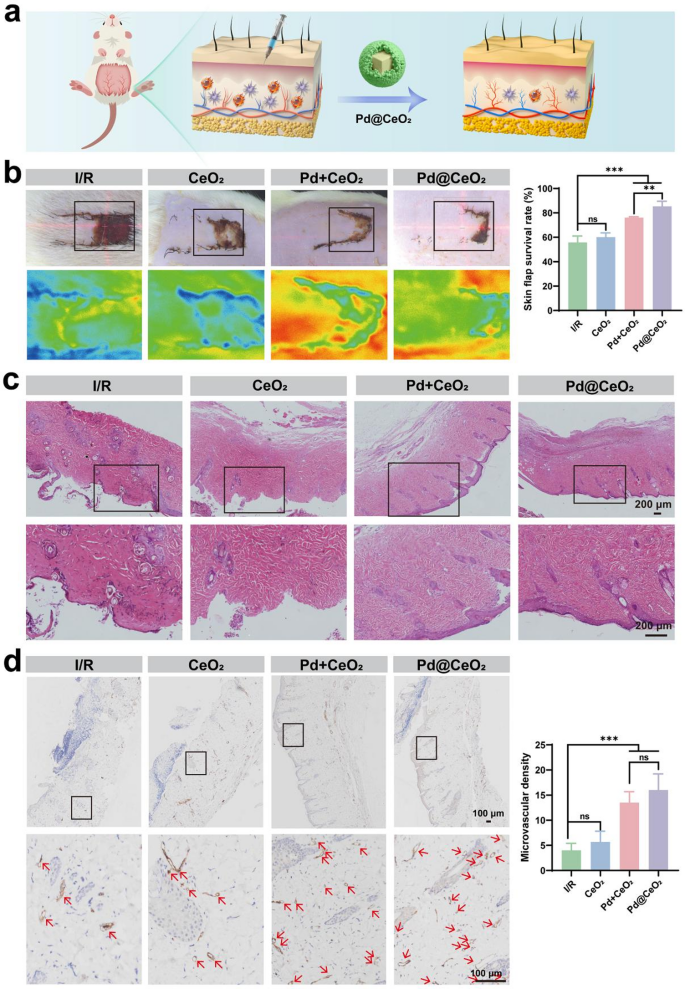
Evaluation of the therapeutic effects of Pd@CeO2 in a rat skin flap I/R injury model. (a) Schematic illustration of the therapeutic process in the I/R injury model. (b) Representative images of skin flaps on day 7 post-surgery for the I/R, CeO2, Pd + CeO2, and Pd@CeO2. (c) HE staining of distal skin flap tissue sections. Scale bars: 200 μm. (d) Representative images of immunohistochemical staining of CD31 in I/R, CeO2, Pd + CeO2, and Pd@CeO2 groups
Mitigation of inflammation and apoptosis by Pd@CeO2 in I/R skin flaps
To further validate the anti-inflammatory potential of Pd@CeO2, immunohistochemical staining of CD68 was conducted to quantify macrophage infiltration, indicative of inflammation within skin flaps. As shown in Fig. 8a, markedly elevated macrophage densities were detected in both the I/R and saline groups (≈ 11.17 each), reflecting pronounced inflammatory activation. In contrast, macrophage infiltration was substantially reduced in the CeO2 (≈ 6.0) and Pd + CeO2 groups (≈ 4.17), with the most pronounced attenuation observed in the Pd@CeO2-treated group (≈ 3.5), thereby underscoring the potent in vivo anti-inflammatory efficacy of Pd@CeO2. Additionally, TUNEL assays were employed to evaluate apoptosis within the flap tissues. Distinctly higher densities of TUNEL-positive cells were detected in the I/R and saline control groups, reflecting significant apoptotic activity (apoptosis rates of 92.67% and 95.67%, respectively). Treatment groups showed substantially reduced apoptosis rates, with CeO2 at 45.68%, Pd + CeO2 at 44.49%, and notably, Pd@CeO2 demonstrating the lowest apoptosis at 43.08% (Fig. 8b). Finally, DHE staining indicated that Pd@CeO2 treatment effectively suppressed ROS accumulation in flap tissues, reinforcing the correlation between robust ROS scavenging and improved flap viability and structural integrity (Fig. 8c). These results collectively confirm that Pd@CeO2 effectively mitigates apoptosis, potentially via robust ROS scavenging and reduced inflammatory signaling, thus underscoring its substantial therapeutic potential in addressing oxidative and inflammatory damage during I/R injury.
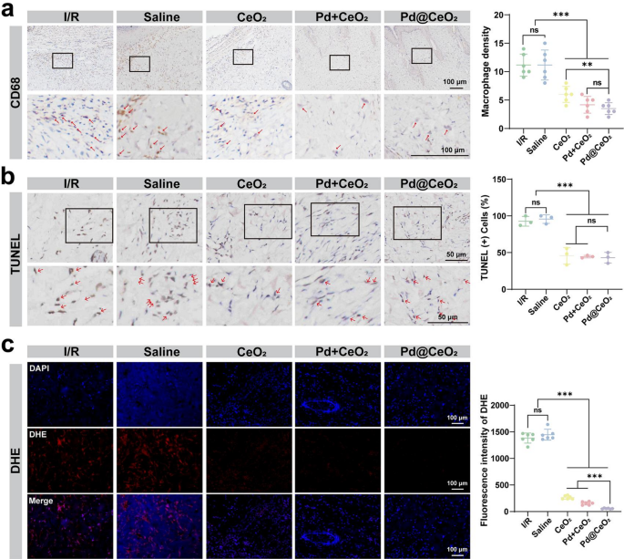
Anti-inflammatory and anti-apoptotic effects of Pd@CeO2 in an I/R skin-flap model. (a) Immunohistochemical staining and semi-quantitative analysis of CD68 to evaluate macrophage densities in the skin flap tissues, with red arrows indicating macrophages, bar = 100 μm. (b) TUNEL staining and quantitative assessment to detect apoptotic cells in skin flap tissues, with red arrows highlighting apoptotic cells. Scale bars: 50 μm. (c) ROS levels and semi-quantitative analysis in skin flap tissues assessed via DHE staining. Scale bars: 100 μm
Transcriptomic analysis of Pd@CeO2-mediated protection against I/R injury
To gain comprehensive insights into the molecular mechanisms underlying the therapeutic actions of Pd@CeO2 in I/R injury, transcriptomic profiling was conducted. Hierarchical clustering of differentially expressed genes (DEGs) revealed distinct transcriptional signatures among the Control, I/R, and Pd@CeO2-treated groups (Fig. 9a). Volcano plots further delineated significantly altered gene expressions, highlighting 2004 upregulated and 2167 downregulated genes when comparing the control and I/R groups. In contrast, Pd@CeO2 treatment induced significant expression changes, with 269 genes upregulated and 138 genes downregulated compared to the I/R group (Fig. 9b). Gene Ontology (GO) enrichment analysis indicated that Pd@CeO2 treatment notably influenced biological processes related to inflammation and immune responses, cellular components associated with membrane and extracellular matrix, and molecular functions involving receptor activity and signaling transduction (Fig. 9c). Notably, enriched biological processes such as “inflammatory response”, “immune response”, and “cellular response to cytokine stimulus” were significantly modulated upon Pd@CeO2 treatment, indicating its robust anti-inflammatory potential at the transcriptomic level. Kyoto Encyclopedia of Genes and Genomes (KEGG) pathway analysis further highlighted key signaling pathways significantly regulated by Pd@CeO2 treatment, including TNF, Chemokine, and PPAR signaling pathways (Fig. 9d). These results corroborate earlier findings regarding the involvement of these pathways in inflammatory modulation and tissue repair, providing robust transcriptomic validation for the anti-inflammatory and regenerative effects observed in previous cellular and histological assessments.
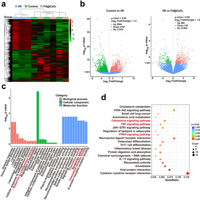
In vivo transcriptomic analysis of rat with I/R injury. (a) Hierarchical clustering heatmap depicting DEG in different groups. (b) Volcano plots. (c) GO enrichment analysis. (d) KEGG pathway enrichment scatterplot


